Table of Contents
ToggleA dark spot on a dental X-ray does not automatically mean a drill. Dentists sometimes look at a small area of decay, check it against your history and risk factors, and decide the smartest move is to watch it instead of filling it. This is called arrested decay, and it happens when a cavity has stopped progressing and no longer poses an active threat to the tooth.
Dentists deliberately leave some cavities untreated when the lesion has stalled in enamel, shows no active breakdown, and the patient’s oral environment supports remineralization instead of continued decay. This is not neglect. It is a documented clinical strategy backed by evidence on nonrestorative caries management.
Not every spot that looks like a cavity behaves like one. Understanding the difference between active and arrested decay changes how you think about your own checkups, and why a dentist at a practice like Castro Valley Advantage Dental might say “let’s keep an eye on that” instead of scheduling a filling.
When Can a Dentist Monitor Decay Instead of Filling It?
A dentist can monitor decay instead of filling it when the lesion is confined to enamel, shows no signs of active breakdown, and the tooth’s surrounding conditions favor repair over destruction. This distinction rests on whether the decay process is still moving or has effectively stopped.
What Arrested Decay Means
Arrested decay is a cavity that has stopped progressing, often described clinically as a lesion that behaves like a scar rather than an open wound. The demineralization process that created the spot has halted, usually because fluoride exposure, saliva, and improved hygiene shifted the balance back toward tooth repair.
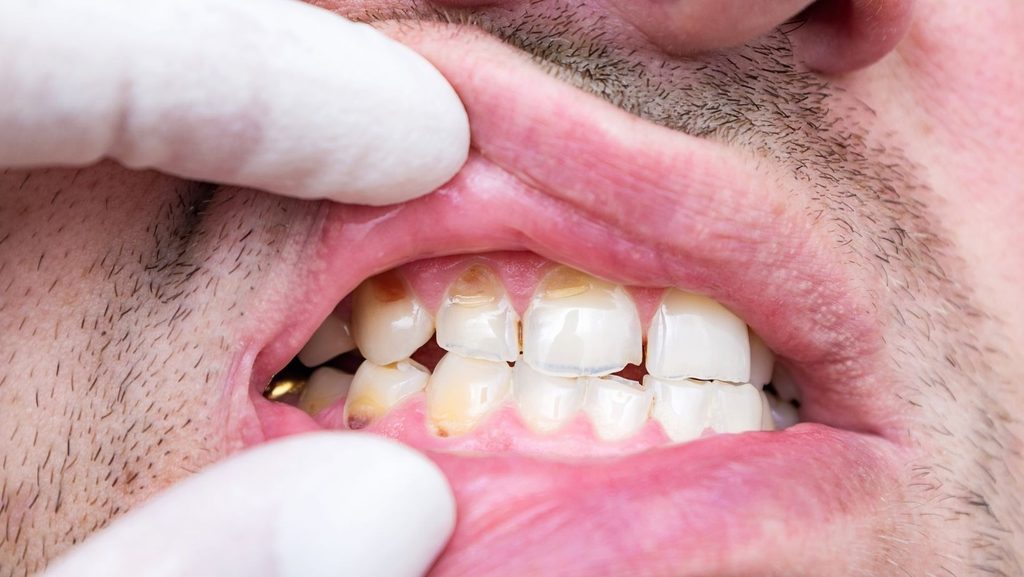
These lesions often appear as dark, hard, shiny spots rather than soft, chalky, or discolored ones. The tooth structure remains intact even though a visible mark exists.
Why Not Every Early Lesion Is an Active Cavity
Early tooth decay does not always mean irreversible damage. The National Institute of Dental and Craniofacial Research notes that early-stage decay can sometimes reverse before it ever becomes a true hole in the tooth.
A white or brown spot on enamel can represent mineral loss that has not yet broken through the surface. At this stage, the tooth can still rebuild itself with the right conditions.
Clinical Signs That a Lesion Has Stopped Progressing
Dentists look for specific physical markers before calling a lesion arrested:
- Hard, glassy, or polished surface texture instead of soft or sticky
- Dark brown or black color rather than light chalky white (a sign of active demineralization)
- No cavitation, meaning no visible break in the enamel surface
- Stable size and appearance across multiple visits, confirmed with X-ray comparison over time
A lesion that meets these markers consistently across repeat exams is a strong candidate for continued observation.
How Dentists Decide Whether Observation Is Safe
Dentists decide observation is safe by combining what they see in the exam chair, what X-rays confirm about depth and location, and what they know about a patient’s daily habits and risk factors. No single factor makes this call alone.
Exam Findings, X-Rays, and Cavity Location
A visual and tactile exam tells a dentist whether a lesion is soft (active) or hard (arrested). X-rays add a second layer of information, showing whether decay has stayed within enamel or extended toward dentin.
Location matters. A cavity in a pit or fissure behaves differently than one on a smooth surface or between teeth. Lesions confined to enamel, with X-rays showing no progression over a defined re-evaluation period (often up to a year for enamel-only decay), are the ones most likely to be cleared for monitoring rather than immediate treatment.
The Role of Fluoride, Saliva, Diet, and Home Care
Fluoride exposure strengthens enamel and helps push the mineral balance toward repair. Saliva flow matters too, since saliva buffers acid and delivers minerals back to weakened enamel.
Diet plays a direct role: frequent sugar or acid exposure keeps a lesion active, while reduced snacking gives enamel time to recover between meals. A patient who brushes with fluoride toothpaste, keeps up with cleanings, and controls diet gives a monitored lesion a real chance to stay arrested.
Why Patient Risk Level Changes the Recommendation
A low-risk patient with good home care and no history of rapid decay is a better candidate for watchful waiting than someone with multiple active cavities or poor plaque control. Dentists weigh caries risk assessment alongside the lesion itself.
The same-looking spot might be monitored in one patient and filled in another, based entirely on that person’s broader risk profile.
When Monitoring Is Not the Right Choice
Monitoring stops being appropriate the moment a lesion shows signs of active progression, cavitation, or symptoms like pain or sensitivity. At that point, observation carries real risk instead of being a safe, conservative option.
Warning Signs of Active or Deepening Decay
Active decay tends to look soft, sticky, and chalky white or light brown rather than dark and hard. Growth between visits, confirmed by X-ray comparison, is a clear signal that a lesion has moved past the arrested stage.
A cavity that once looked stable but now shows a rough or broken surface texture has likely resumed progressing and needs treatment rather than continued observation.
Pain, Sensitivity, and Infection Risks
Pain is an unreliable early warning system. Sensitivity to cold, sweets, or pressure can signal that decay has reached dentin, and by the time discomfort shows up, the lesion may already be past the point where monitoring makes sense.
Decay that progresses past enamel and reaches the nerve can cause significant pain and may require a root canal or extraction instead of a straightforward filling. Ignoring escalating symptoms turns a simple restoration into a much larger procedure.
Why a Hole in Dentin Usually Needs Treatment
Once decay creates an actual cavitation into dentin, the tooth can no longer repair itself the way it can in early enamel lesions. Dentin is softer and more vulnerable to bacteria, and a physical hole gives decay a direct path deeper into the tooth.
At this stage, a filling, inlay, onlay, or more advanced restoration becomes necessary depending on how far decay has spread. Waiting past this point raises the risk of infection, nerve involvement, and eventual tooth loss.
Protecting Teeth Through Informed Monitoring
Arrested decay reflects a real, evidence-supported approach to cavity management, not a shortcut or a missed diagnosis. The decision to monitor instead of fill depends on lesion appearance, X-ray findings, location, and each patient’s individual risk factors.
Regular checkups are what make this strategy safe. A dentist can only confirm that a lesion has stayed stable, rather than started progressing again, by comparing exams and X-rays over time.
If you have a spot your dentist is watching, keep every follow-up appointment and stay consistent with fluoride use and home care. That consistency is what keeps a monitored lesion arrested instead of letting it turn into a cavity that needs a filling, a root canal, or worse.