
Table of Contents
ToggleTwo dentists can stare at the same bitewing and walk away with opposite plans because reading an X-ray is a judgment call, not a measurement. The image shows shadows of density and shape; deciding what those shadows mean, and what to do about them, depends on training, thresholds, and the clinical picture around the tooth. Research on diagnostic agreement among dentists backs this up: agreement rates on the same radiographs range widely, with disagreement showing up in 20% to over 50% of cases depending on the finding in question.
That gap unsettles patients more than almost any other part of dental care. You expect an X-ray to be black-and-white evidence, and instead you get two different treatment plans from two licensed professionals looking at the identical film.
Disagreement between dentists over the same X-ray usually reflects differences in clinical judgment, risk tolerance, and image quality, not a mistake by either provider. Knowing where that judgment comes from gives you a way to evaluate both opinions, and to ask better questions of a dental team like Galleria Dentistry, instead of guessing which dentist to trust.
What Different Readings of the Same X-Ray Can Mean
Two different readings of one X-ray can mean several things at once: a difference in what each dentist sees, a difference in how each dentist would act on what they see, or both. Untangling which one you’re dealing with is the first step to making sense of conflicting advice.
Diagnosis and Treatment Are Separate Decisions
A diagnosis identifies what’s present on the tooth; a treatment plan decides what to do about it. Two dentists can agree completely on the diagnosis, such as early enamel decay on a molar, and still disagree on treatment, with one recommending a filling now and the other recommending monitoring at the next visit. When you compare opinions, separate these two layers instead of treating “different plan” as proof of “different diagnosis.”
Why Disagreement Does Not Automatically Signal Poor Care
Variation in dental opinions is a documented, normal feature of clinical practice, not evidence that one dentist is incompetent or dishonest. Dentistry operates with clinical guidelines rather than rigid rules, and reasonable practitioners applying those guidelines to the same borderline finding can land in different places. A second opinion that differs from the first is routine, not a red flag by itself.
Where Interpretation Differences Are Most Common
Interpretation gaps show up most in borderline, early-stage findings rather than obvious disease. Early interproximal decay, faint periapical shadows, and early bone loss around the gumline are the classic gray zones. Advanced decay, a fractured root, or a visible abscess rarely produce disagreement; the image is unambiguous at that stage.
Why Dental Opinions Vary
Dental opinions vary because five factors stack on top of each other: personal thresholds for treatment, the technical limits of the X-ray itself, what the dentist finds in your mouth beyond the image, differences in training and philosophy, and your individual history and risk profile. Each factor alone can shift a recommendation; together they explain most of the gap between two treatment plans.
Different Thresholds for Watching or Treating Early Decay
Some dentists treat early decay the moment it appears on film; others prefer to monitor it and intervene only if it progresses. Neither approach is wrong on its own. A dentist who fills small lesions early argues it prevents larger, costlier problems later. A dentist who watches argues early lesions can remineralize or stay stable for years, and treating them adds unnecessary drilling. Patients on Reddit dental forums describe this exact split: “some dentists are more conservative and like to watch smaller areas of decay vs immediately filling them.”
X-Ray Quality, Angle, and Limits of the Image
The physical image itself introduces variation before any human judgment enters the picture. A cone-beam CT scan reveals detail a two-dimensional bitewing cannot; an older X-ray machine produces lower resolution than a newer digital sensor. Even the angle of the shot changes what’s visible, since overlapping teeth or a slightly off-axis image can hide or exaggerate a shadow that looks like decay. Research on radiograph interpretation confirms that image type, whether bitewing, panoramic, or CBCT, directly affects diagnostic accuracy and the rate of interpretation errors.
Clinical Findings Beyond the X-Ray
An X-ray is one input among several a dentist uses to reach a conclusion. A visual exam, a probe test for softness, patient-reported sensitivity, and bite function all inform the diagnosis, and two dentists who weigh these inputs differently can reach different conclusions from the same image. A dentist who examines your mouth in person and finds soft, sticky enamel might treat something the X-ray alone made look borderline.
Training, Experience, and Treatment Philosophy
Dental school, continuing education, and years of practice shape how a dentist reads ambiguous findings. Some practitioners lean toward early, preventive intervention; others favor a watch-and-wait approach grounded in evidence that early lesions can be managed non-surgically. Neither philosophy is the industry default. Both are represented in published clinical guidance.
Your Risk Factors and Dental History
Your cavity history, oral hygiene habits, and overall risk profile change how a dentist interprets the same finding. A borderline spot on the X-ray of a patient with a history of rapid decay progression gets treated more aggressively than the identical spot on a patient with strong hygiene and no cavity history in a decade. Your dentist’s recommendation is partly about you, not only about the image.
How to Compare Two Treatment Recommendations
Comparing two treatment plans starts with asking each dentist to point to the specific finding driving their recommendation, then lining up the goal, urgency, and alternatives side by side. This turns a confusing disagreement into a concrete comparison you can evaluate.
Ask Each Dentist to Show You the Finding
Request that each dentist point directly to the spot on the X-ray or intraoral camera image that justifies the recommended treatment. A dentist who can show you a specific shadow, dark spot, or bone-level change is working from a visible finding. A dentist who struggles to point to anything concrete may be recommending based on a lower threshold for treatment rather than a clear-cut finding.
Compare the Goal, Urgency, and Alternatives
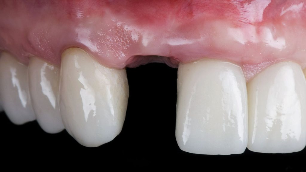
Two different-sounding plans sometimes lead to the same outcome. One dentist suggesting a dental implant and another suggesting a bridge are both solving the same missing-tooth problem through different methods. Ask each dentist:
- What is this procedure meant to fix, health or appearance?
- What happens if I wait six months and get a re-check instead?
- What are the alternative options, and why did you rule them out?
| Comparison Point | Plan A | Plan B |
| Primary goal | Function/health | Function/health |
| Urgency | Immediate | Watch and recheck |
| Alternative considered | None mentioned | Remineralization/monitoring |
| Cost | $ | $ |
Request Your X-Rays and Clinical Records
You’re entitled to copies of your own X-rays and clinical notes, and getting them makes an independent comparison possible. Ask the first office for digital copies of the film and written notes before your second appointment. This avoids a second dentist re-taking X-rays unnecessarily and lets any reviewer compare the identical image both original dentists saw.
When an Independent Second Opinion Makes Sense
A second opinion makes sense when the treatment is costly, irreversible, or urgent, such as a root canal, extraction, or full crown, and the first recommendation doesn’t match your symptoms. It’s also worth pursuing when a new dentist finds several new problems a previous provider never mentioned. Bring your existing X-rays to the second visit rather than starting from a blank record, and ask the second dentist to review the same image before forming an opinion.
Making a Confident Decision With the Evidence Available
You don’t need a tie-breaking third opinion to move forward with confidence. Focus on whether each dentist can point to a specific, visible finding, whether the proposed treatment matches the urgency of that finding, and whether the risks of waiting have been explained in plain terms.
Two reasonable dentists reading the same X-ray differently is a known, documented pattern in dentistry, not a sign that something has gone wrong. Treat a second opinion as useful data rather than a verdict on the first dentist’s competence. Ask for the specific finding, compare the goal and urgency of each plan, and request your own records so any reviewer works from the same image. That process, more than the disagreement itself, is what gets you to a decision you can trust.